The 2023 Nobel Prize in Physiology or Medicine has been awarded to Katalin Karikó and Drew Weissman for their breakthrough discoveries to use modified RNA for therapeutic treatments. Together with Herbert Tschochner, we have written a short comment to celebrate the occasion. We highlight the importance of the discoveries made by Katalin Karikó and Drew Weissman and how they have contributed to the development of RNA-based therapies. The comment can be accessed at Pflugers Archiv – European Journal of Physiology via this link.
RNA interactome studies suggested that numerous metabolic enzymes possess the ability to bind RNA. This has fueled the REM hypothesis that describes the regulatory interplay of RNAs, metabolic enzymes and metabolites. In a collaborative effort, a consortium comprising the Meister, Suess, and Rossbach labs and headed by the Babinger lab, has investigated RNA binding of select enzymes in E.coli using iCLIP, SELEX, MST, and EMSA experiments. This revealed specific RNA interaction of glutamate-5-kinase (ProB) and quinone oxidoreductase (QorA). It will be exciting to learn about the functional consequences and the biological importance of these protein RNA interactions. The open-access publication can be found here: weblink.
In a close collaboration with Monica Pichler from the lab of Thomas Mock at at he University of East Anglia, we could establish ribosome profiling for the Diatom Thalassiosira pseudonana. This unicellular organism belongs to an important group of eukaryotic microalgea that play a key role as primary producers in aquatic ecosystems, generating 20-50% of the oxygen on the planet each year, and comprising approx. half of the organic material found in the oceans. With ribosome profiling now available for diatoms, it can now be determined how changing environmental conditions such as acidification of the oceans or an increase in water temperature affect protein synthesis in the model organism T.pseudonana. Please find the open access publication at Current Protocols: weblink.
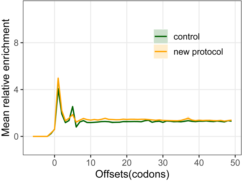
Our protocol for rapid ribosome profiling of small input samples has been published in Nucleic Acids Research. We demonstrate the outstanding performance of the newly developed sequencing library preparation workflow and its reproducibility with minute amounts of sample (0.1 pmol of RNA). We would like to thank our collaborators (Meister lab at the University of Regensburg, Leidel lab at the University of Bern & König lab at IMB Mainz) for their support! Please find the open access publication here: weblink.
Are you interested in establishing ribosome profiling in your lab to analyze cellular translation comprehensively and quantitatively?
Check out our novel and simplified protocol! The standardized workflow employs an extremely rapid sequencing library preparation protocol (12 hours) that relies on solid phase extraction of reaction intermediates, making it easy to implement in any standard laboratory. The protocol yields data of extremely high quality from minute amounts input material allowing the analysis of samples that were previously not easily amenable to this type of experimentation.
Our study of the activity of tha TRIM-NHL protein Mei-P26, a regulator of cell fate in Drosophila, has now been published at Life Science Alliance (doi 10.26508/lsa.202201418).
In a close collaboration with the labs of Sebastian Glatt (Max Planck Research Group at the Malopolska Centre of Biotechnology, Jagiellonian University Krakow, Poland) and Janusz Bujnicki (Laboratory of Bioinformatics and Protein Engineering, International Institute of Molecular and Cell Biology in Warsaw, Poland), we could solve the first high resolution structure of the Mei-P26 NHL domain and define a consensus RNA motif that it recognizes. Molecular dynamics simulations allowed us to predict and subsequently experimentally validate key amino acid residues involved specific RNA recognition, highlighting differences to other NHL domains. Using individual nucleotide resolution cross-linking and immunoprecipitation (iCLIP), we could identify RNA targets of Mei-P26 in cultured Drosophila cells and demonstrate the protein can either repress or activate its genuine mRNA targets. Regulation requires the NHL domain of the protein but is independent of its function as a ubiquitin ligase.
In particular, the last finding significantly expands our understanding of TRIM-NHL protein-mediated gene regulation. These proteins were previously considered to exclusively act as repressors of gene expression. Strikingly, Mei-P26 itself appears to lack any regulatory activity suggesting that the regulatory outcome is determined by the recruitment of different co-factors, some of which have previously been identified.
Pünktlich zum Weltkrebstag, dem 04. Februar, erscheint eine Pressemitteilung über unsere Forschungsergebnisse. Gemeinsam mit unseren Partnern Robert Ahrends, Grischa Tödt, Björn Tews und Christiane Knobbe-Thomsen, haben wir einen neuen Mechanismus entdeckt, der in Tumorzellen eine Resistenz gegen weit verbreitete Chemotherapeutika auslöst. Diese Chemoresistenz hat dramatische Folgen für betroffene Patienten, da die Behandlung stark eingeschränkt wird. Ein besseres Verständnis des Mechanismus der Chemoresistenz soll zukünftig neue Therapieoptionen ermöglichen.
The TRIM-NHL protein Meiotic-P26 acts as a regulator of cell fate in Drosophila. Its activity is critical for ovarian germline stem cell maintenance, differentiation of oocytes and spermatogenesis. Together with our collaborators from the Glatt lab (Max Planck Research Group at the Malopolska Centre of Biotechnology, Jagiellonian University Krakow, Poland) and the Bujnicki lab (Laboratory of Bioinformatics and Protein Engineering, International Institute of Molecular and Cell Biology in Warsaw, Poland), we could solve the first high resolution structure of the Mei-P26 NHL domain and define a consensus RNA motif that it recognizes. Molecular dynamics simulations allowed us to predict and subsequently experimentally validate key amino acid residues involved specific RNA recognition, highlighting differences to other NHL domains. Using individual nucleotide resolution cross-linking and immunoprecipitation (iCLIP), we could identify RNA targets of Mei-P26 in cultured Drosophila cells and demonstrate the protein can either repress or activate its genuine mRNA targets. Regulation requires the NHL domain of the protein but is independent of its function as a ubiquitin ligase.
In particular, the last finding significantly expands our understanding of TRIM-NHL protein-mediated gene regulation. These proteins were previously considered to exclusively act as repressors of gene expression. Strikingly, Mei-P26 itself appears to lack any regulatory activity suggesting that the regulatory outcome is determined by the recruitment of different co-factors, some of which have previously been identified by genetic means.
A preprint of the manuscript is available at bioRxiv (doi.org/10.1101/2021.09.20.461029)
On Tuesday, July 20th Andreas Horn very successfully defended his PhD thesis. For many years, Andreas was advancing our understanding of how RNA binding proteins control development and cell fate decisions in the model organism Drosophila melanogaster. We are very proud and happy about Andreas’ success and we wish him all the best for his future endeavors.
Are you excited about biomedical research? Do you want to employ state-of-the-art methodology with the aim to unerstand how tumor cells become resistant to treatment? Do you want to work in a stimulating research environment with great colleagues? Then this is the right job for you:
We are looking for a Technical Assistant to perform ribosome profiling experiments in the context of cellular stress and resistance to chemotherapeutic treatment. Click here for more information(PDF, german).
Please feel free to contact us any time for more details…
A combination of high-throughput analyses uncovers novel mechanism of stress-induced chemoresistance
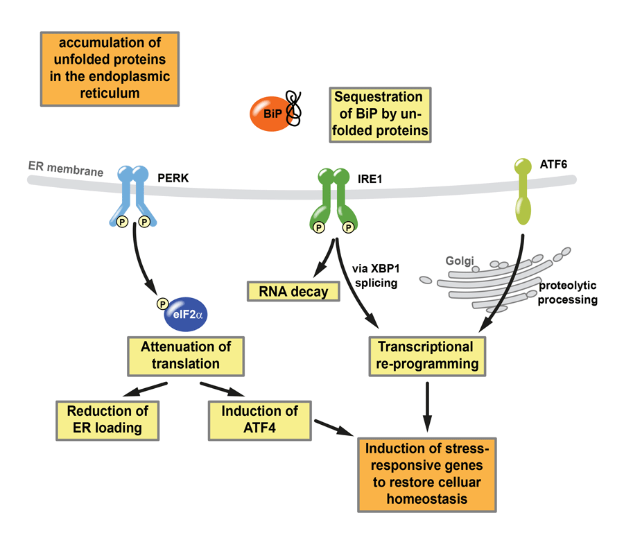
Resistance of cancer cells against therapeutic agents is a major cause of treatment failure, especially in recurrent diseases. In a collaborative effort with the labs of Robert Ahrends, Björn Tews, Grischa Tödt and Christiane Knobbe-Thomsen, we identified a novel mechanism of chemoresistance which has now been published in ‘Nature Communications’. It is driven by the Unfolded Protein Response (UPR), a cellular stress response pathway that alters gene expression and cellular metabolism to promote cell survival under stress.
The Unfolded Protein Response (UPR), an important cellular stress response pathway, does not only contribute to cancer development, progression and chemoresistance, but also it plays an important role in numerous other diseases, among them diabetes and neurodegenerative disorders such as Alzheimer’s disease. A detailed biochemical understanding of the UPR is critically required to better define its role in disease and to develop novel therapeutic strategies. To produce a comprehensive description of the UPR, we employed a ‘multi-omics’ approach, combining large datasets from genetics and proteomics. This allowed us to define a list of genes (the UPR regulon) that are activated to promote cell survival under stress. Besides the previously known factors, we identified numerous genes that have not previously been implicated in stress response pathways and many of them have key functions in cancer development and cellular metabolism.
Schematic overview over the Unfolded Protein Response (UPR)
Changes in cellular metabolism are a hallmark of cancer cells and allow to sustain rapid tumor growth. Chemotherapy often aims at interfering with these metabolic pathways. We demonstrated that stress-mediated genetic regulation of enzymes involved in amino acid biosynthesis and one-carbon (1C) metabolism that relies on the vitamin folate as a cofactor. Moreover, upon stress, cancer cells become fully resistant to chemotherapeutic agents which target this specific metabolic pathway. This includes Methotrexate, a drug commonly employed in the treatment of cancer and rheumatic disease. Detailed biochemical and genetic investigations revealed that resistance is driven by a previously unrecognized mechanism. Its precise molecular characterization might lead to novel therapeutic concepts aimed at overcoming chemoresistance n cancer therapy.
Publication:
Reich S, Nguyen CDL, Has C, Steltgens S, Soni H, Coman C, Freyberg M, Bichler A, Seifert N, Conrad D, Knobbe-Thomsen CB, Tews B, Toedt G, Ahrends R, and Medenbach J: A multi-omics analysis reveals the unfolded protein response regulon and stress-induced resistance to folate-based antimetabolites – in Nature Communications, DOI:10.1038/s41467-020-16747-y
A function in splicing versus a function in translational control – why not both?
U2AF proteins are best known for their functions in spliceosomal processing of pre-mRNAs where a homodimer of U2AF65 and U2AF35 functions in recognition of the 3′ splice site. The smaller subunit, U2AF35, contains two zinc fingers (ZnFs). Mutations therein have recently been associated with malignant transformation. The molecular function(s) of the two domains have, however, not been studied in great detail.
A collaborative effort, spearheaded by the Heyd lab at the Free University of Berlin, now revealed that the two ZnFs have remarkably different activity. Both are required for splicing regulation, whereas only ZnF2 controls protein stability and contributes to the interaction with U2AF65.
Intriguingly, a naturally occuring splice variant of U2AF26, a paralog of U2AF35, lacks the second ZnF. It is upregulated upon activation of primary mouse T cells and localizes to the cytoplasm, suggesting a splicing-independent function. Employing ribosome profiling in a model T cell line, we provide evidence for a role of U2AF26 in activating cytoplasmic steps in gene expression, notably translation. Consistently, an MS2 tethering assay shows that cytoplasmic U2AF26/35 increases translation when localized to the 5ʹUTR of a model mRNA. This regulation is partially dependent on ZnF1 thus providing a connection between a core splicing factor, the ZnF domains and the regulation of translation. Altogether, our work reveals unexpected functions of U2AF26/35 in mammalian cells beyond the regulation of splicing.
Andreas Meindl has just joined the lab as a PhD student. Andreas will work on the function of Sex-lethal in Drosophila melanogaster sexual development addressing in detail its auto-regulatory feedback to splicing. Welcome to the lab!
You want to work on exciting and diverse research projects employing state-of-the-art methodologies? You want to join a young and highly motivated team? Then get your CVs ready, there is a job opening for a Technical Assistant!
The basic information: Salary TV-L E9, starting date as soon as possible, application deadline December 22nd 2019.
For more information (in German) please click here.